News
Soldiers exposed to bomb blasts may be at higher risk of Alzheimer’s

Soldiers exposed to the shock waves from military explosives may be at an increased risk of developing Alzheimer’s in later life.
A study supported by the Leonard Wood Institute in Missouri in cooperation with the US Army Research Laboratory (ARL), has found that the brains of otherwise healthy military personnel who are exposed to explosions show an abnormal accumulation of amyloid-beta protein.
The build-up of certain forms of this toxic protein is known to cause brain cells to become sick and die, leading to cognitive decline and neurodegenerative diseases, such as Alzheimer’s.
Research conducted over several decades has suggested there may be a relationship between repetitive or severe traumatic brain injury (TBI) caused by contact sports like rugby and football, assaults, falls, and traffic accidents, and abnormal amyloid-beta accumulation.
TBI can also result from indirect forces, however, such as shock waves from battlefield explosions, that shake the brain violently in the skull.
Previous autopsy studies have shown the presence of amyloid plaques as early as hours after severe brain injury.
For this latest study published in Radiology, a journal of the Radiological Society of North America, researchers recruited nine military grenade or breacher instructors at Fort Leonard Wood Military Base in Fort Leonard, Missouri, from January 2020 to December 2021.
Grenade and breacher instructors are military officers who train recruits in the use of hand grenades and explosives or other mechanical methods to force open doors.
An additional nine civilians were included in the study as a healthy control group. All participants had no previous history of concussion, and they were all males in their early 30s, an age at which amyloid accumulation is not expected.
The 18 participants were evaluated twice. The first evaluation was to establish a baseline and the second occurred after blast exposure, approximately five months after the baseline examination.
The military instructors filled out a digital log with the number of exposures to explosions, including the firing of weapons. The control participants were evaluated at similar time points. 
All participants underwent a PET scan of the head to evaluate and quantify amyloid changes. Analysis software was used to segment six brain regions that are usually associated with Alzheimer’s disease and TBI.
Abnormal amyloid accumulation was seen in six of the nine participants who were exposed to explosions. Three of the participants had one region of the brain with increased amyloid accumulation, two had two areas, and one had three zones with abnormal accumulation.
None of the healthy control participants showed any abnormal amyloid build-up.
Study author Carlos Leiva-Salinas, associate professor of radiology at the University of Missouri School of Medicine in Columbia, Missouri, said: “Amyloid-beta is a molecule not normally found in the brains of young patients. Amyloid-beta accumulation in the brain is proposed to be an early event in the pathogenesis of Alzheimer’s disease, the most common type of dementia worldwide, impacting millions of people.
“Further research needs to be done to establish the relationship between the frequency and the severity of traumatic brain injury and the degree of amyloid changes in the brain, the natural course of the observed accumulation, and other potential biologic risk factors for amyloid plaque deposition and the development of cognitive decline.”
He suggested that non-invasive positron emission tomography, or PET, imaging could be used to identify early-stage amyloid-beta accumulation in individuals or professions exposed to traumatic brain injury such as military personnel, police officers, firefighters, and sports people, like football players.
This is not the first study to make a link between soldiers exposed to explosions and a higher risk of Alzheimer’s.
Analysis published in February 2021 conducted by researchers at the University of North Carolina in collaboration with the US Army Combat Capabilities Development Command, the ARL, and the National Institutes of Health, that tested the impact of controlled military blast waves on rats, identified selective reductions in components of brain connections that are required for memory.
The researchers also observed a sharp drop in electrical activity from those neuronal connections. They stated that blast-induced effects were evident among healthy neurons, which could explain the increased risk of Alzheimer’s disease among soldiers without any apparent brain damage.
At the time the ARL’s Army Research Office programme manager, Dr Frederick Gregory, said: “Blasts can lead to debilitating neurological and psychological damage but the underlying injury mechanisms are not well understood.
“Understanding the molecular pathophysiology of blast-induced brain injury and potential impacts on long-term brain health is extremely important to understand in order to protect the lifelong health and wellbeing of our service members.”


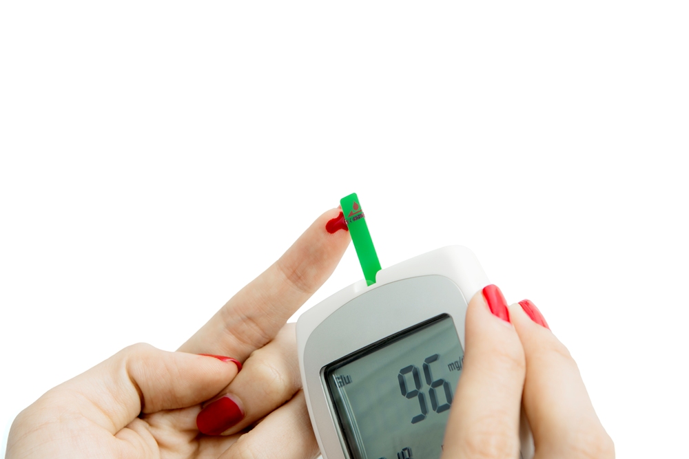
Sharp rises in blood sugar after meals may raise Alzheimer’s risk, according to genetic analysis of more than 350,000 adults.
The findings point to after-meal glucose, rather than overall blood sugar, as a possible factor in long-term brain health.
Researchers examined genetic and health data from over 350,000 UK Biobank participants aged 40 to 69, focusing on fasting glucose, insulin, and blood sugar measured two hours after eating.
The team used Mendelian randomisation, a genetic method that helps test whether biological traits may play a direct role in disease risk.
People with higher after-meal glucose had a 69 per cent higher risk of Alzheimer’s disease.
This pattern, known as postprandial hyperglycaemia (elevated blood sugar after eating), stood out as a key factor.
The increased risk was not explained by overall brain shrinkage (atrophy) or white matter damage, suggesting after-meal glucose may affect the brain through other pathways not yet fully understood.
Dr Andrew Mason, lead author, said: “This finding could help shape future prevention strategies, highlighting the importance of managing blood sugar not just overall, but specifically after meals.”
Dr Vicky Garfield, senior author, added: “We first need to replicate these results in other populations and ancestries to confirm the link and better understand the underlying biology.
“If validated, the study could pave the way for new approaches to reduce dementia risk in people with diabetes.”

Shingles vaccine may slow biological ageing in older adults

Thousands of men in England to be offered life-extending prostate cancer drug

Blood sugar spike after meals may increase Alzheimer’s risk

Study reveals why memory declines with age

Agetech research round-up: brain health vital, £38m to combat Alzheimers, and more…

FDA clears automated brain fluid device

Agetech World’s latest innovation & investment round-up

Insilico signs US$888m oncology deal with Servier

Food preservatives linked to increased diabetes and cancer risk, study finds

UK bans junk food ads before 9pm to protect child health
 News2 weeks ago
FDA clears automated brain fluid device
- News2 weeks ago
Agetech World’s latest innovation & investment round-up
- News2 weeks ago
Insilico signs US$888m oncology deal with Servier
- Markets & Industry2 weeks ago
Food preservatives linked to increased diabetes and cancer risk, study finds
- News2 weeks ago
UK bans junk food ads before 9pm to protect child health
- News1 week ago
Captioning glasses win AARP pitch at CES
- Insights2 weeks ago
Global longevity initiative launches North American chapter
- News6 days ago
Interview: GlycanAge launch first hospital-based tests